

It is the sixth leading cause of cancer death in the United States. Most of these tumours are superficial with a median overall survival rate of over 90%, but with a high recurrence rate. Its basic treatment is transurethral resection of the bladder and intravesical instillations, depending on the histology of the tumour. In cases of infiltrating tumour, it is necessary to apply more aggressive treatments such as radical cystectomy or external radiation therapy.
In our Lyx Centre for Urology in Madrid we will perform all the necessary techniques for diagnosis and treatment, as well as close collaboration, in case needed, with the Medical Oncology, Radiation Oncology, Pain Unit services, for complementary treatments (chemotherapy, radiation therapy, etc.).
The available procedures and techniques are the following:
- Techniques for diagnosis and endourological treatment (cystoscopy, exploration under anesthesia, ureterorenoscopy diagnosis, transurethral resection of the bladder, Holmium-Yag Laser Fulguration on a same-day surgery basis)
- Ambulatory intravesical instillations of chemotherapy or immunotherapy (mitomycin C, BCG, etc.)
- Advanced Surgical Techniques
- Open radical cystectomy, laparoscopic or assisted by robot, with extended lymphadenectomy.
- Non-continent or continent urinary diversion using orthotopic bladder substitution
- Open, laparoscopic or robot-assisted nephroureterectomy
What is the bladder?
The urinary bladder is a hollow muscular organ that stores urine before later emptying. Urine is produced in the kidneys and reaches the bladder through tubes called ureters. The emptying of the bladder is performed through ducts called urethra. The muscle of the bladder (detrusor) helps the emptying of the bladder via contraction. A thin layer, called the urothelium, lines the inside of the bladder. Immediately after, there is an area of connective tissue called the lamina propria. Surrounding the lamina propria, the bladder muscle is covered in turn by fat as well as by a surface called the peritoneum.
¿How do you develop Bladder Cancer?
Currently, we only understand part of the origin and progression of bladder cancer. However, we have identified substances that may foster the development of bladder cancer. The most important are related to tobacco. It is estimated that smoking alone is responsible for 50% of all cases of bladder cancer. Chronic exposure to certain substances, primarily those contained in paints and solvents, cause another 20% to 25% of these cases.
The transformation of normal cells into cancerous cells originates in the urothelium. 90% of the bladder tumours originate in this place. Most of the tumours that we diagnose have not progressed beyond this urothelium and the next layer (i.e. the lamina propria) and have not invaded the muscle layer yet. These are the so-called muscle-infiltrating tumours of the bladder. Although they are malignant tumours, their location gives them the characteristic of NOT being invasive (i.e. they have not spread to the muscle layer).
Most can be treated without need to remove the bladder. The tumours that invade the muscle layer (called muscle-invasive tumours) are generally more aggressive in nature and represent a greater risk, with less therapeutic options. These tumours usually require a more aggressive approach (such as the radical surgery of the bladder, when it is necessary to extract the entire organ).
What are the main symptoms of bladder cancer?
The most common symptom is Hematuria (blood in the urine), which is usually monosymptomatic. Eventually, the entire bladder cancer will present blood in the urine. In some cases, blood can be seen in the urine during urination (hematuria), while in other cases, the blood is only observed under a microscope (microscopic hematuria) and is detected after completing a General Examination of Urine. The presence of hematuria does not in itself confirm the existence of bladder cancer. There are many other causes of blood in the urine. For example, it may be a result of a urinary tract infection. Once hematuria is diagnosed, it is necessary to carry out further studies to determine whether it is bladder cancer or not.
How is Bladder Cancer diagnosed?
 The initial medical assessment with a complete clinical history and physical examination. We will ask you if you have a risk factor for bladder cancer such as smoking or exposure to chemical agents. In addition, as the hematuria may come from any part of the urinary tract (kidneys, ureters, bladder, prostate, urethra), we need to conduct studies to rule out the presence of problems at these levels. Special urine tests can provide us with important information. We can search for suspicious cells with a microscope, which would guide the diagnosis of bladder cancer, urine cytology.
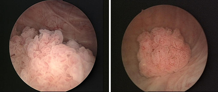
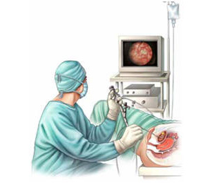
The most important diagnostic test is cystoscopy, which allows to observe under direct vision the inside the bladder. This procedure may be performed in our CUMQ BERNABEU STADIUM centre and consists in introducing a special lens called a cystoscope through the urethra into the inside of the bladder.
If a tumour is found, it is important to note the size, location, and appearance before its resection (removal). This procedure is performed with another instrument called a resectoscope, which is a similar to the cystoscope lens, but it has a handle at the end to resect the tumour. This procedure is called transurethral resection of bladder tumour (TURBT). The resected tissue is then sent to a pathologist for analysis.
What are the options for treating non-muscle invasive bladder cancer?
The treatment of choice for patients with tumours confined to the urothelium (Ta) and/or to the lamina propria (T1) consists in the transurethral resection of bladder tumour (TURBT). There are other alternative treatments that are comparable with the RTU-V, such as laser therapy; however, the RTU-V has certain advantages such as obtaining tissue to be sent to the pathologist to determine the grade and stage of the tumour. With the laser, the structure of the tumour remains extremely distorted and usually, it is difficult to obtain tissue for analysis.
Intravesical chemotherapy and immunotherapy
After resection, chemotherapy or intravesical immunotherapy can be used to prevent the recurrence of tumours. The term “intravesical” refers to “within the bladder”, so it involves placing the drugs directly inside the organ. The main agents currently used are mitomycin, and Bacillus Calmette- Guérin (BCG).
[maxbutton id=”13″ url=”https://www.lyxurologia.com/en/uro-oncology/bladder-cancer/treatment-of-bladder-cancer-with-chemohyperthermia/” text=”Treatment of Chemohyperthermia” ]
Radical cystectomy
Cystectomy (surgical removal of the bladder) may be an option in patients with carcinoma in situ (CIS) or T1 tumours of the high degree that persists or has recurred after an initial intravesical treatment. There is a high risk of progression to muscle-invasive tumour in these patients and some may consider the option of radical cystectomy as a first choice of treatment. If so, we will discuss the information in relation to the risks of the procedure, as well as to the method of bladder reconstruction.
What are the options of treating muscle-invasive bladder cancer?
In the cases of muscle-invasive bladder cancer, the treatment has to be radical. The first treatment option is radical cystectomy (removal of the bladder) preceded or not by treatment with chemotherapy, depending on the case. Once the bladder removed, urinary diversion (reconstruction using the bowel for the output of urine) is performed within the same procedure.